
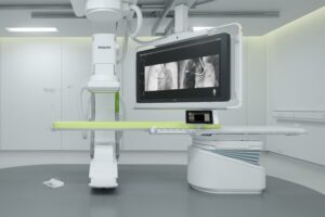
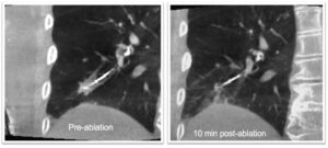
Philips introduced Azurion Lung Edition, an advanced 3D imaging and navigation platform, to accelerate lung cancer diagnosis and treatment. The suite combines CT-like 3D images acquired at the tableside (Cone Beam CT) with live X-ray guidance and advanced tools to support image-guided lung procedures. The system is designed specifically for bronchoscopy procedures, enabling clinicians to perform both minimally invasive endobronchial biopsies and lesion ablation during the same procedure. By speeding diagnosis and treatment, Azurion Lung Edition helps transform lung cancer care, contributing to dramatically improved outcomes and reduced costs.

Each year 1.7 million people die worldwide of lung cancer, making it the leading cause of cancer deaths globally [1] and accounting for more loss of life than breast, colon, and prostate cancer combined. Today, over 60% of patients are diagnosed at a late stage, with a small chance of a surgical cure. But thanks to a rise in the number of lung cancer screening programs and increasing patient awareness, a growing proportion of patients have small peripheral lesions that are operable.

In addition to early diagnosis, fast treatment is critical to ensure better outcomes and quality of life for lung cancer patients, with every week of delay resulting in a 5% increase in mortality. Today, most patients face a long journey to a definitive diagnosis, and they often undergo a painful recovery after open surgery. Cone Beam CT is seen as the gold standard for clinicians to diagnose and treat lung cancer in one room, and even during the same procedure.
“This is a very exciting time in the world of interventional pulmonology and advanced bronchoscopy,” said Michael Pritchett, pulmonary and critical care physician, director, Chest Center of the Carolinas, FirstHealth Moore Regional Hospital, Pinehurst Medical Clinic, US. “One of the things that we’re particularly excited about is being able to diagnose patients, stage their cancer and treat them, all in a single procedure. As a diagnostic bronchoscopist it’s exciting and rewarding to be able not only to diagnose patients, but to go on to treat them as well.”
“Lung cancer is one of the most aggressive forms of cancer, and we need innovative technology to fight back,” said Gustavo Cumbo-Nacheli, director of Bronchoscopy and Interventional Pulmonology at Spectrum Health in Grand Rapids, Michigan, US. “Without a Cone Beam CT scan to confirm placement of the biopsy needle, repeat procedures are often necessary. And by combining Cone Beam CT with other technologies, including robotics, we will be able to go beyond biopsy and treat the patient.”
On Wednesday, September 23, at 13:00 ET (19:00 CET). Atul Gupta, interventional radiologist and chief medical officer, Philips Image Guided Therapy, will host an exclusive virtual roundtable together with a panel of renowned pulmonologists and interventional radiologists, “The Future Now – Implementing Cone Beam CT-guided endobronchial ablation therapy”. To register, click here.
“Image-guided minimally invasive procedures continue to expand into new treatment areas, enabled by sophisticated, procedure-oriented solutions like Azurion Lung Edition,” said Gupta. “With lung cancer increasingly being detected at an earlier stage, new minimally invasive treatment strategies like ablation have the potential to significantly improve outcomes for patients.”
Intuitive, integrated, and efficient, the Azurion platform optimizes clinical and operational lab performance and expands the role of image-guided interventions in treating patients. The platform has achieved rapid global adoption and has been used in well over two million procedures worldwide.


