
On 19 May 2021, Royal Philips, a global leader in health technology, announced its newest solution for precision diagnosis with the global introduction of its spectral detector-based Spectral Computed Tomography (CT) 7500. This latest intelligent system delivers high-quality spectral images for every patient on every scan 100% of the time to help improve disease characterization and reduce rescans and follow-ups, all at the same dose levels as conventional scans.

The time-saving spectral workflow is fully integrated, enabling the technologist to get the patient on and off the table quickly – spectral chest scans and head scans take less than one second, and a full upper body spectral scan can be completed in less than two seconds – while still delivering high-quality imaging that allows the physician to rapidly deliver a confident diagnosis and effective treatment plan for each patient.

The human and financial cost of misdiagnosis
Missed and delayed diagnoses contribute to roughly 10% of patient deaths annually [1], while an estimated 10-20% of all medical diagnoses are inaccurate [2]. The financial costs resulting from unnecessary, suboptimal, and repeat imaging costs as much as US$ 12 billion annually [3]. The new Spectral CT 7500 was designed for first-time-right diagnosis and has demonstrated a 34% reduction in time to diagnosis, a 25% reduction in repeat scans, and a 30% reduction in follow-up scans [4].
With such great human and financial costs due to misdiagnosis, Spectral CT 7500 sets a new standard of care where image quality, dose, and workflow come together to deliver valuable clinical insights.
“With such great human and financial costs due to misdiagnosis, Spectral CT 7500 sets a new standard of care where image quality, dose, and workflow come together to deliver valuable clinical insights. This latest intelligent system helps to bring clarity to defining moments in healthcare by delivering on certainty, simplicity, and reliability in every clinical area from cardiac care to emergency radiology, diagnostic oncology, intervention, and radiation oncology,” said Kees Wesdorp, chief business leader of Precision Diagnosis at Philips. “Our detector-based spectral technology ensures spectral data is always available and is seamlessly integrated into current workflows, meaning scans are fast, and clinicians are able to send patients the right treatment pathway with a more confident diagnosis.”
Spectral CT 7500 has improved scanning procedure
Spectral CT has demonstrated higher sensitivity in detecting malignant findings and has improved readings of incidental findings [5] [6]. With Philips spectral detector CT, photons add more value by helping salvage sub-optimal injection scans without the need to re-scan the patients, shortening the time to diagnosis.
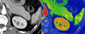
“We rely on CT scans to provide us valuable insights. But conventional CT scanners are limited and can only show us where things are located – like lesions, cysts, bleeds, fractures and more. Philips spectral detector-based systems help to characterize what the finding is, not just where it is, providing us greater confidence in diagnoses,” said Dr. Finn Rasmussen, associate professor, consultant Radiologist, MD and MSc. at Aarhus University. “We have seen significant reductions in rescans and follow-ups by adopting spectral into our workflow for faster and more accurate diagnosis [7].”
Expanded patient populations and optimized workflows
Spectral CT 7500 expands on Philips proven spectral-detector benefits to now include additional patient populations that were not previously served. The spectral insights are available for all patients, from pediatric to bariatric, and for any clinical indication, including challenging cardiac scans with high and irregular heart rates, without compromising image quality, dose, or workflow. The spectral workflow enables radiologists to optimize reading with rich spectral results and AI-based smart tools available in any reading environment with Spectral Magic Glass on PACS.
[1] https://pubmed.ncbi.nlm.nih.gov/23011708/
[2] https://www.coverys.com/About-Us/Foundation/Grants-For-Improving-Diagnostic-Accuracy
[3] https://reactiondata.com/wp-content/uploads/2015/02/peer60:unnecessaryimaging.pdf
[4] Analysis by CARTI Cancer Center in Little Rock Arkansas and University Hospitals of Cleveland – Results from case studies are not predictive of results in other cases. Results in other cases may vary.
[5] Analysis by Aarhus University Hospital Aarhus, Denmark. Results from case studies are not predictive of results in other cases. Results in other cases may vary.
[6] Analysis by University Hospital Cleveland, USA. Results from case studies are not predictive of results in other cases. Results in other cases may vary.
[7] Results from case studies are not predictive of results in other cases. Results in other cases may vary.


