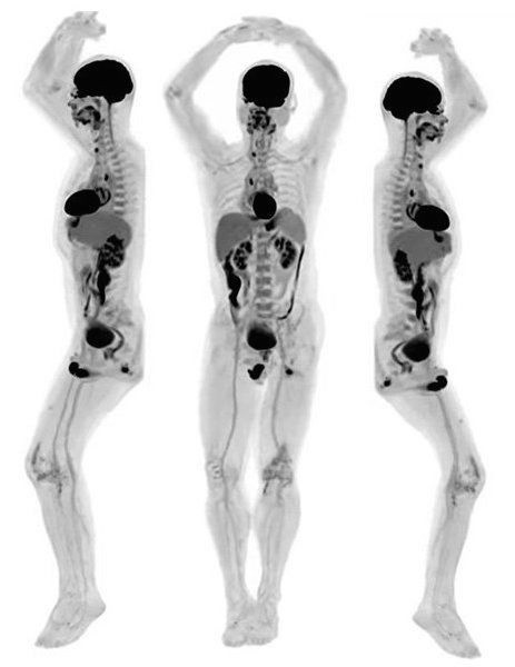
A positron emission tomography (PET) scan is a nuclear medicine functional imaging technique that allows doctors to observe metabolic processes in the body and aids in the diagnosis of diseases. The scan uses a special dye containing radioactive tracers. These tracers are either swallowed, inhaled, or injected, depending on what part of the body is being examined.
Scientists at the University of California Davis (UCD) professor Simon Cherry, department of biomedical engineering, and Ramsey Badawi, chief of nuclear medicine at UCD Health and vice-chair for research in the department of radiology, have successfully developed a scanner for total body scan. Cherry and Badawi conceived this idea almost 15 years ago, and it was kickstarted in 2011 with support from the National Cancer Institute and the National Institutes of Health.
Explorer, the world’s first total-body imaging scanner, can capture a 3D picture of the whole human body at once. It has the capability to scan up to 40 times faster, and it uses up to 40 times less radiation dose than current PET systems. Because it captures radiation more efficiently than other scanners, it can produce an image in as little as one second and can create a diagnostic scan of the whole body in as little as 20 seconds.
Explorer is a combined positron emission tomography (PET), and X-ray computed tomography (CT) scanner that can image the entire body at the same time. It can also capture real-time videos of blood flow and heart function. According to developers, the technology has countless applications, from improving diagnostics to tracking disease progression to researching new drug therapies. Recently, researchers have published their findings in the Proceedings of the National Academy of Sciences journal in January 2020.
Tracking changes in real-time
According to Qi and project scientist Xuezhu Zhang, the combination of the scanner and advanced data reconstruction enables real-time tracking of blood flow over the human circulatory system, motion-frozen heart beating and breathing monitoring for cardiovascular and cerebrovascular disease and analysis of respiratory system function. Zhang has developed methods to reduce noise and reconstruct images from the raw data from Explorer scans of volunteers. They were able to see changes on a scale of 100 milliseconds or one-tenth of a second and use these to create high-quality real-time movies of their scans.
Unique features of Explorer
According to the developers, the quality of images is much better and allows physicians to see smaller tumors and other diseases in advance. The scanner can be set up to run much faster than conventional PET scanners, which can make it easier and more comfortable for patients. This is particularly good for young children or patients with joint pain. The scanner can alternatively be set up to use much less radiation, which is helpful for children, or research where the same person needs to be scanned many times. The entire body can be imaged at the same time, which means it can track changes in a drug’s distribution throughout the whole body, enabling an understanding of the concentration of medicine in every organ and tissue over time.
The scanner is now commercially available from United Imaging Healthcare, Shanghai.


