")
Royal Philips, a global leader in health technology, and Radboud university medical center (Radboudumc), a leading Dutch academic research and teaching hospital (Nijmegen, the Netherlands), has announced the positive results of a clinical study conducted by Radboudumc aimed at setting a new standard of safety and accuracy in the diagnosis of small peripheral lung lesions. The study outcomes represent a promising milestone in the global innovation effort to set a new clinical standard for detecting lung cancer at an early stage, potentially leading to improved prognoses for patients.

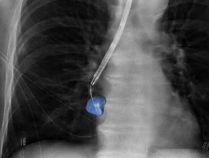
Radboudumc’s observational study reported the diagnostic accuracy and procedural radiation dose for patients undergoing an endobronchial lung biopsy supported by Philips Lung Suite – a solution that uses 3D imaging with augmented fluoroscopy to support high precision diagnosis and minimally-invasive therapy in one room. Diagnostic accuracy of 90% was reported while reducing the average total effective radiation dose per procedure by more than half from 47.5 Gy·cm2 (effective dose: 14.3 mSv) to 25.4 Gy·cm2 (effective dose: 5.8 mSv). The median long-axis diameter of the 248 lesions navigated to during the study was 13 mm (range 5-65mm).

Lung cancer – the leading cause of cancer death
Lung cancer kills around 1.7 million people a year worldwide. That makes it the leading cause of cancer death globally, accounting for greater loss of life than breast, colon, and prostate cancer combined. While early diagnosis and treatment are critical to better outcomes and quality of life, the majority of lung cancers are currently diagnosed at a late stage, with minimal chance of a surgical cure.
Philips Lung Suite enables all-in-one lung cancer diagnosis and treatment. It provides advanced real-time 3D imaging with augmented fluoroscopy on the company’s Image-Guided Therapy System – Azurion, combined with dedicated software. With Philips’ Cone Beam CT imaging, the X-ray detector rotates around the patient to generate a CT-like image in around five seconds, providing clinicians with a high-resolution 3D view of the target lesion and other anatomical structures. This allows the clinician performing the biopsy procedure to be continually guided by high-quality real-time imaging to advance a catheter towards the lesion through a bronchoscope. Once done, its position can be confirmed in real-time using the same imaging modality and a biopsy sample removed.
Philips has a comprehensive portfolio of lung cancer diagnosis and treatment solutions. In addition to Philips Lung Suite, the company’s Lung Cancer Orchestrator provides an integrated lung cancer patient management system for both CT lung screening programs and incidental pulmonary findings programs that manage and monitors patients every step of the way in their lung cancer screening and treatment decision journey.


